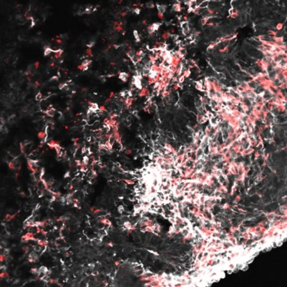

Generation Process
Through our proprietary differentiation technology, hPSC-derived midbrain organoids can be cultured long term and develop into advanced models enriched with mature dopamine neurons. These organoids offer a reliable system for investigating neurodevelopment and neurodegenerative diseases, including Parkinson’s.

Cellular and Structural Similarity
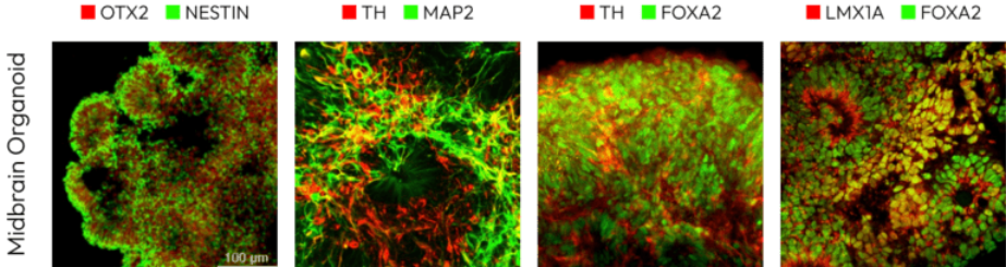
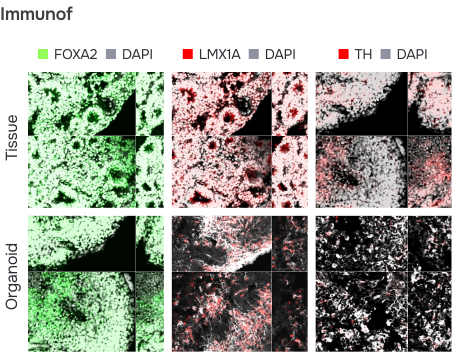
Our midbrain organoids replicate the key genetic and structural features of the human midbrain. They contain abundant dopamine neurons along with diverse supporting cell types. When cultured long term, they even develop melanin in the substantia nigra – a hallmark feature unique to the human midbrain.
FOXA2 – A transcription factor crucial for the development and differentiation of midbrain and hindbrain neurons, promoting the formation of midbrain dopaminergic neurons.
LMX1A – A transcription factor that regulates the differentiation of dopaminergic neurons in the midbrain and hindbrain.
TH (Tyrosine Hydroxylase) – An enzyme involved in dopamine synthesis, commonly used as a marker for dopaminergic neurons.
Nestin – A marker for neural progenitor cells and immature neurons, indicating immature cells in midbrain organoids.
OTX2 – A transcription factor important for the development of the forebrain and midbrain, expressed in the midbrain and brainstem regions.
Functional similarity
Parkinson’s Disease Modeling
We can generate organoid models of neurodegenerative diseases through two methods: creating brain disease organoids using iPSCs with regulated expression of specific genes or inducing brain disease organoids through drug treatment.
Two main approaches are used to model Parkinson’s disease. The first involves utilizing gene editing technologies to modify or alter genes associated with Parkinson’s, such as introducing mutations in LRRK2 or SNCA to replicate disease-causing genes and analyze pathological features. The second approach involves creating organoids from patient-derived iPSCs (induced pluripotent stem cells).
By using iPSCs from Parkinson’s patients, personalized disease models can be developed, allowing researchers to experimentally recreate disease characteristics like dopaminergic neuronal degeneration and study drug development and therapeutic strategies.

Result
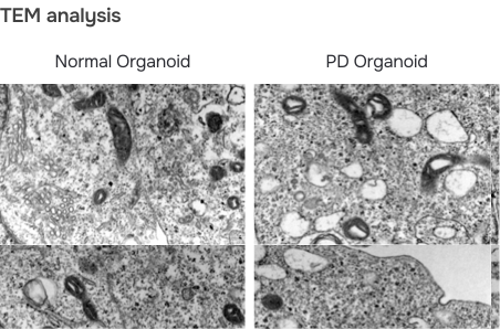
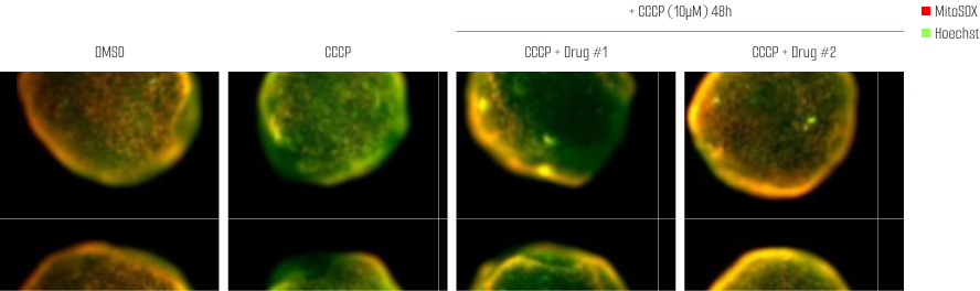
Parkinson’s disease is marked by the progressive loss of dopamine-producing neurons, driven in part by mitochondrial dysfunction. Mitochondrial swelling – a pathological enlargement of mitochondria – disrupts energy production and calcium balance, leading to cell death. This dysfunction also heightens oxidative stress and impairs intracellular signaling, ultimately damaging dopamine neurons. Together, these processes drive the motor symptoms of Parkinson’s disease, including tremors and movement difficulties.
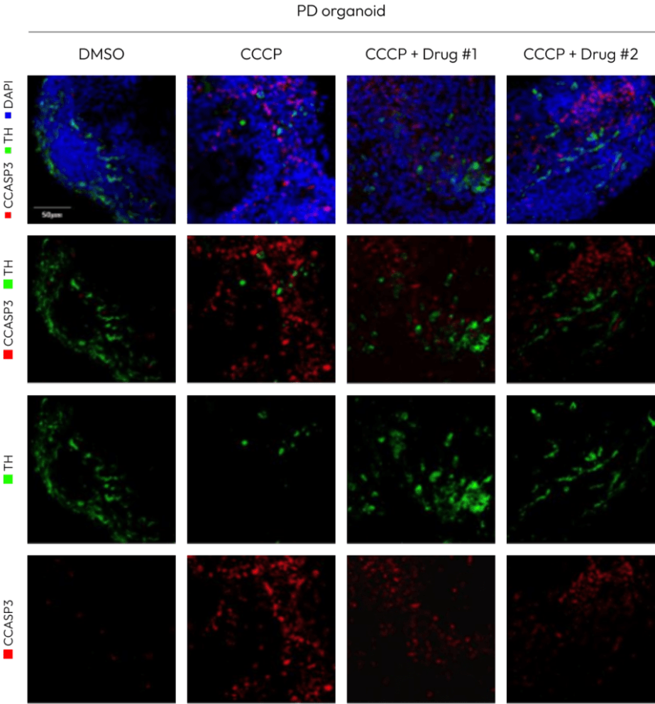
The experiment comparing the expression of th (tyrosine hydroxylase) and ccasp3 (cleaved caspase-3) after treatment with CCCP (a mitochondrial toxin) in Parkinson’s disease organoids is crucial for studying mitochondrial dysfunction and cell death mechanisms.
CCCP depolarizes the mitochondrial membrane potential, inhibiting mitochondrial function and inducing damage to dopamine-producing neurons. In the experiment, th serves as a marker for dopamine-producing neurons, while ccasp3 is a marker for apoptosis.
The results showed a decrease in th expression and an increase in ccasp3 expression following CCCP treatment. This suggests that mitochondrial dysfunction induced by CCCP leads to damage to dopamine neurons and enhances cell death. These findings help confirm the link between mitochondrial dysfunction and cell death pathways in Parkinson’s disease and may offer valuable insights for developing new therapeutic approaches.
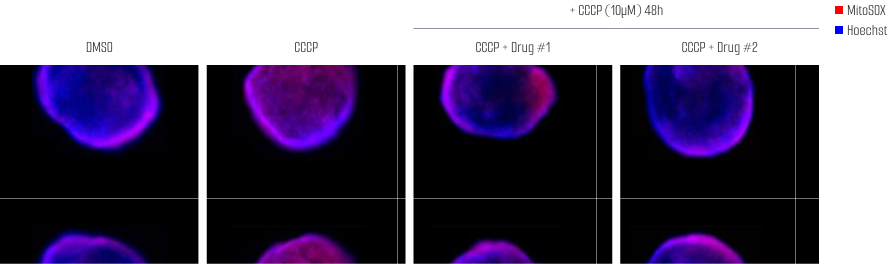
MitoSOX (ROS indicator)
JC-1 (Mitochondrial membrane potential