Label-Free Lipid Quantification Using Holotomography
Holotomography is ideal for studying lipid metabolism in living cells, allowing time-lapse observation without staining or phototoxicity. It effectively detects high refractive index molecules like lipid droplets, making it valuable in cell biology, bioengineering, and biofuels.
- Label-free, non-invasive lipid quantification
- Live-cell compatible for longitudinal studies
- 3D quantitative analysis of lipid droplets
- No staining artifacts or phototoxicity
Applications
- Lipid metabolism research
- Metabolic disease models
- Drug screening for lipid accumulation
- Long-term live-cell imaging
Read out
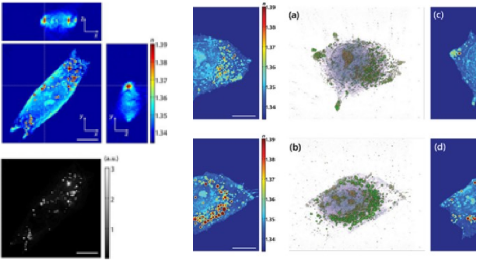
Holotomography of a human hepatocyte, Huh-7
3D tomogram images (Left) fluorescence stained with Nile Red (Right). Reproduced by from Kim et al. Scientific Reports 6:36815 (2016).
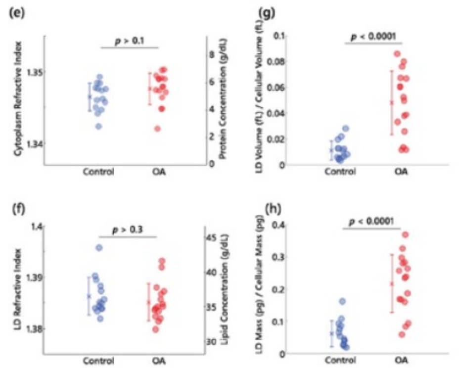
3D RI tomograms of lipid droplets in live hepatocytes
(a,b) 3D rendered isosurface image of 3D RI distribution of (a) an untreated and (b) OA treated hepatocyte.
(c,d) Cross-sectional slice images of 3D RI distribution of (a) the untreated and (b) OA treated hepatocyte.
The scale bars indicate 10μm. e-h, Quantitative analysis of (e) RI of cytoplasm, (f) RI of LDs, (g) the ratio of LD volume to cell volume, (h) the ratio of LD mass to cellular dry mass, Reproduced from Kim et al., Scientific Reports 6:36815 (2016).
(c,d) Cross-sectional slice images of 3D RI distribution of (a) the untreated and (b) OA treated hepatocyte.
The scale bars indicate 10μm. e-h, Quantitative analysis of (e) RI of cytoplasm, (f) RI of LDs, (g) the ratio of LD volume to cell volume, (h) the ratio of LD mass to cellular dry mass, Reproduced from Kim et al., Scientific Reports 6:36815 (2016).