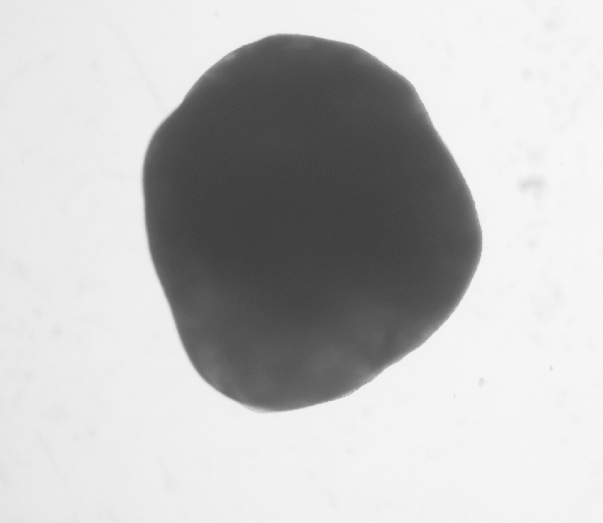
Generation of cortical organoid

Cortical Organoid Cellular and Structural Organization
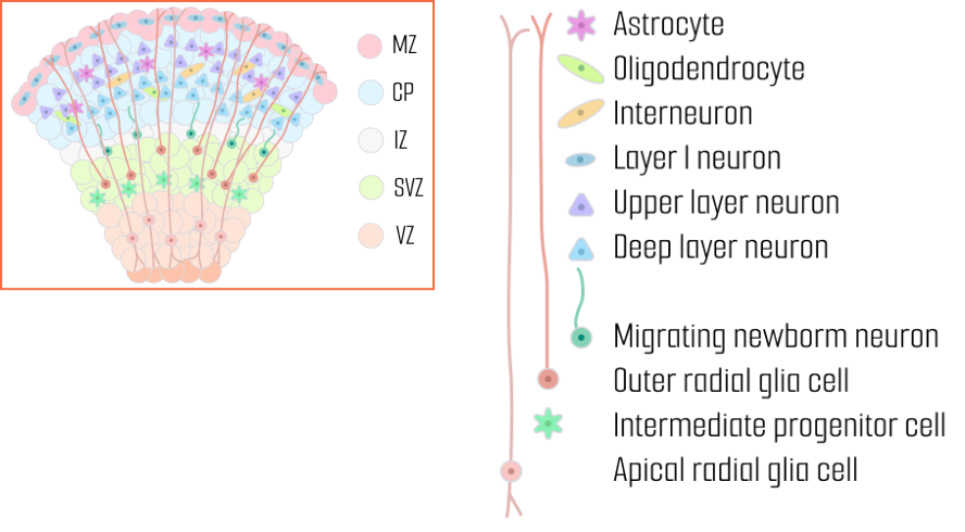
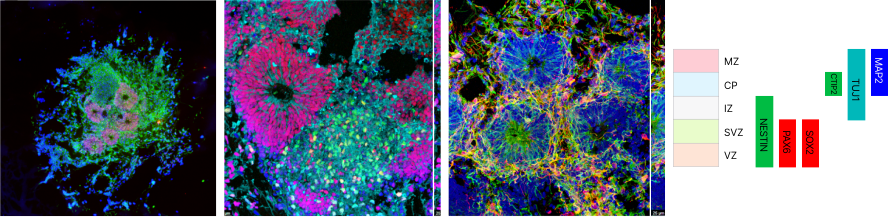
During human cortical development, cortical organoid is organized into five distinct zones
SVZ (Subventricular Zone) – A region where neural progenitor cells differentiate and prepare for migration.
IZ (Intermediate Zone) – The migratory pathway for neurons, where axons and neural fibers are formed.
CP (Cortical Plate) – The region where mature neurons settle to form the cortical structure.
MZ (Marginal Zone) – The outermost layer of the cerebral cortex involved in neural circuit formation and signal modulation.
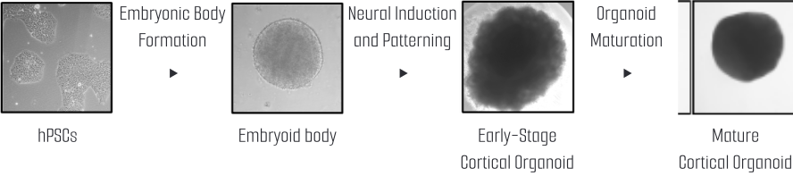
Cortical organoid development from hPSC
Frequency analysis of electrical activities in cortical organoids
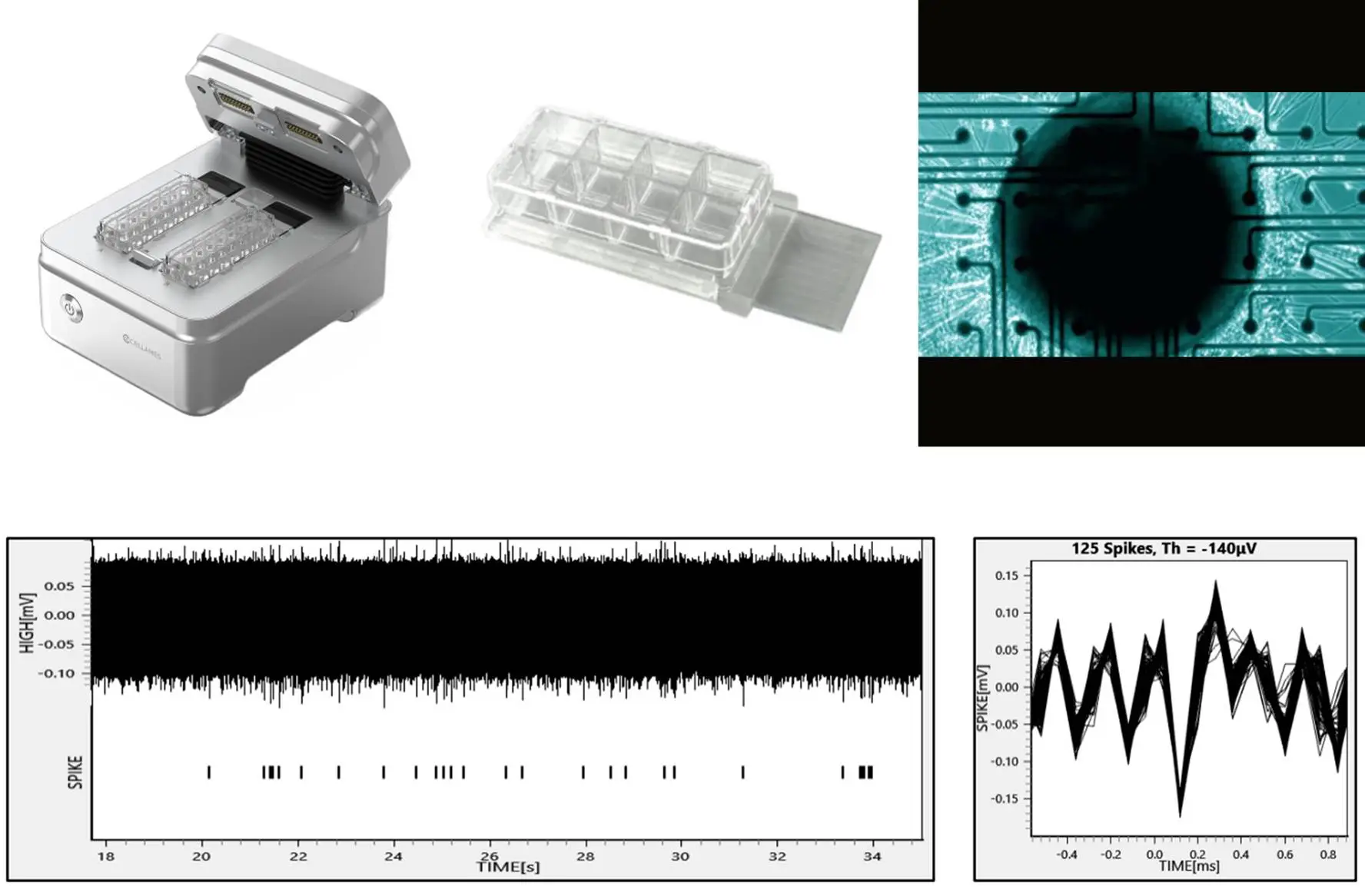
Field Potential Analysis (MEA)
Modeling Brain Diseases with iPSC-Derived Organoids
Scheme for inducing disease in normal brain organoids by drug treatment
Scheme for generating brain organoids using patient-derived induced pluripotent stem cells (iPSCs)
Scheme for generating brain organoids after gene editing via the CRISPR-Cas9 method
Glioblastoma Multiforme (GBM) Models
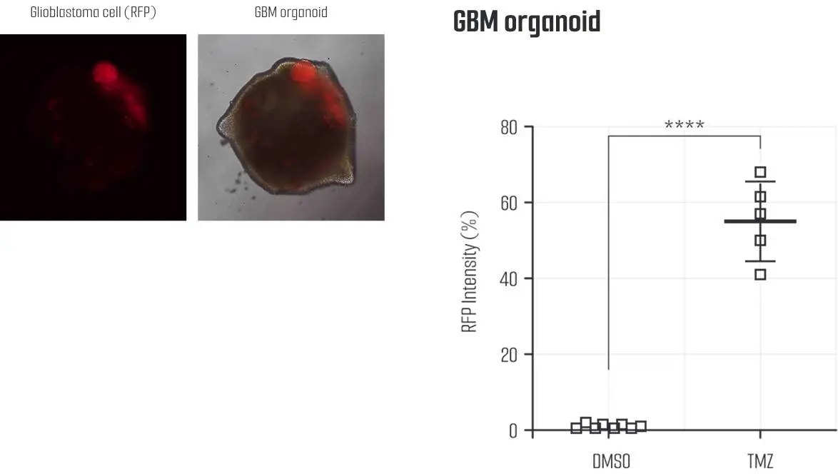
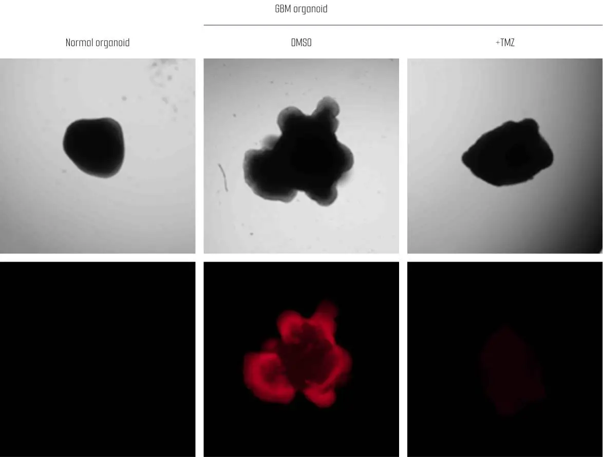
Advance Drug Discovery with Smarter, Ethical Models.
Frequently Asked Questions
What are cortical organoids, and what do they model?
What research applications are cortical organoids most suitable for?
How do Lambda Biologics' cortical organoids enhance research reliability?
What is the difference between cortical organoids and cerebral organoids?
Cortical organoids and cerebral organoids are both brain organoids grown from human pluripotent stem cells (hPSCs), but they are different models.
Cortical organoids are made by guided differentiation. They are specifically patterned to resemble the cerebral cortex and form its layered structure. Because they are highly specific to the cortex, they are highly reproducible with low batch-to-batch variability. They also give better, more consistent results in multi-electrode array (MEA) recordings of electrical activity.
Cerebral organoids are made by unguided differentiation. They form a mix of brain regions, such as cortex-like, retina-like, choroid plexus, midbrain-like and hindbrain-like tissue, and which regions appear depends on the hPSC line used. As a result, their reproducibility is low, batch-to-batch variability is high and MEA results are less consistent.