General Information

Lipid Quantification

Price:
Intracellular lipid accumulation and spatial distribution play a critical role in cellular metabolism, energy homeostasis, and the progression of metabolic and degenerative diseases. However, conventional lipid quantification methods rely on fluorescent dyes or chemical staining, which can perturb cellular physiology, introduce artifacts, and limit long-term or dynamic studies. Lambda Biologics offers label-free lipid quantification using Holotomography (HT) – a non-invasive imaging technology that enables high-resolution, 3D visualization and quantitative analysis of intracellular lipid droplets in live cells, without staining or labeling. By leveraging intrinsic refractive index (RI) contrast, HT preserves the native cellular environment while delivering accurate, reproducible lipid measurements.
Organism
Product Type

Tissue

Disease

Applications

  • Holotomography: Cell biology, LLPS

Label-Free Lipid Quantification Using Holotomography

Holotomography is ideal for studying lipid metabolism in living cells, allowing time-lapse observation without staining or phototoxicity. It effectively detects high refractive index molecules like lipid droplets, making it valuable in cell biology, bioengineering, and biofuels.

  • Label-free, non-invasive lipid quantification
  • Live-cell compatible for longitudinal studies
  • 3D quantitative analysis of lipid droplets
  • No staining artifacts or phototoxicity

Applications

  • Lipid metabolism research
  • Metabolic disease models
  • Drug screening for lipid accumulation
  • Long-term live-cell imaging
A staining-free alternative to traditional lipid dyes, holotomography delivers physiologically relevant, reproducible data for advanced cell-based research.

Read out

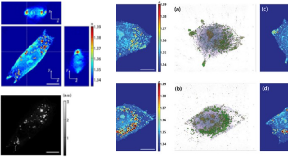
Holotomography of a human hepatocyte, Huh-7
3D tomogram images (Left) fluorescence stained with Nile Red (Right). Reproduced by from Kim et al. Scientific Reports 6:36815 (2016).
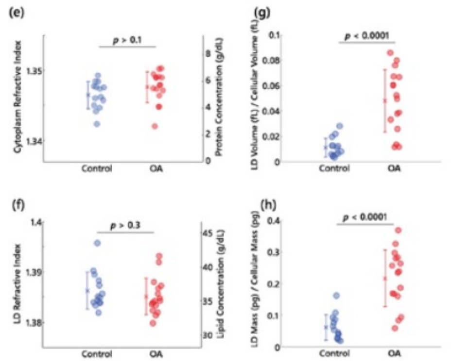
3D RI tomograms of lipid droplets in live hepatocytes
(a,b) 3D rendered isosurface image of 3D RI distribution of (a) an untreated and (b) OA treated hepatocyte.
(c,d) Cross-sectional slice images of 3D RI distribution of (a) the untreated and (b) OA treated hepatocyte.
The scale bars indicate 10μm. e-h, Quantitative analysis of (e) RI of cytoplasm, (f) RI of LDs, (g) the ratio of LD volume to cell volume, (h) the ratio of LD mass to cellular dry mass, Reproduced from Kim et al., Scientific Reports 6:36815 (2016).


Trusted by Leading Innovators

Customer insight

Functional Testing of Anaerobic Microbes Using Organoids

Chong Kun Dang - better life through better medicine

“We have been developing products utilizing various bacterial strains and have explored multiple methods to evaluate the efficacy of our developed strains. However, due to the nature of obligate anaerobic bacteria, there were significant limitations in applying conventional in vitro efficacy evaluation methods. To overcome these challenges, we explored different testing approaches and discovered the ODISEI platform...

Customer insight

Advancing K-Beauty with Skin Organoids

Professor Lee Chang-seok Eulji University

“Advancing K-Beauty with Skin Organoids: A Next-Generation Platform for Non-Animal Testing and High-Precision Cosmetic Innovation With the global rise of K-beauty, the cosmetics industry continues to grow steadily. Since the ban on animal testing for cosmetics in Korea in 2017, various alternative testing methods have been adopted. However, many of these approaches have limitations in replicating the complexity...

Customer insight

HT-X1: A Label-Free Imaging Breakthrough for Organoid-Based Disease Modeling and Drug Screening

Tomocube (Spatial)

“Traditional microscopy methods often require fluorescent labeling to analyze cellular structures, which can be time-consuming and invasive. In contrast, our HT-X1 system allows for high-resolution visualization of cellular morphology without the need for any labeling, offering a clear advantage in live-cell imaging. To validate this capability, we conducted an organoid-based analysis using intestinal organoids...

Customer insight

Advancing Clinical Pathology with Spatial Protein Analysis

Seoul National University College of Medicine

“Traditional protein analysis has primarily focused on quantifying expression levels within tissue samples. However, recent advances in spatial analysis techniques have shifted attention toward evaluating not only expression levels, but also the spatial distribution, density, and proximity between proteins...

Customer insight

Revealing Immune-Boosting Potential with ODISEI-Gut

K Research Institute

“Among the many fermented foods we consume, kimchi is particularly known for containing a diverse range of lactic acid bacteria, which are believed to influence the activation of immune cells in the body. In our research institute, we successfully isolated a bacterial strain that appears to enhance immune cell function. To evaluate its effect in the intestinal environment, we utilized the ODISEI-Gut platform...

Customer insight

Multiplex Marker Analysis Enhances Research Efficiency with 31-Marker Detection on a Single Slide

Bundang Jesaeng General Hospital

“We conducted a study focused on identifying disease-related markers using patient-derived tissue samples. However, traditional methods limited our ability to analyze multiple candidate markers simultaneously, and the limited availability of clinical samples posed a consistent challenge...

Customer insight

Redefining Gastric Cancer Research with Patient-Derived Organoids

SOON CHUN HYANG UNIVERSITY HOSPITAL

“Gastric cancer is one of the leading causes of cancer-related mortality worldwide. It is a malignant solid tumor characterized by poor prognosis due to difficulties in early diagnosis and its rapid progression. Unlike many other solid tumors that primarily metastasize via lymphatic or hematogenous routes, gastric cancer most commonly spreads through the peritoneum....

Customer insight

Breast Cancer PDOs for TNBC Drug Development

GANGNAM SEVERANCE HOSPITAL

“Breast cancer is one of the most commonly diagnosed cancers among women worldwide. It is a complex and heterogeneous disease, with its progression and treatment response varying significantly depending on factors such as hormone receptor status, HER2 expression, and genetic background...

Customer insight

Transforming Gastric Cancer Treatment: Lambda Biologics’ PDOs Platform

SOON CHUN HYANG UNIVERSITY HOSPITAL

“Gastric cancer is one of the leading causes of cancer-related mortality worldwide. It is a malignant solid tumor characterized by poor prognosis due to difficulties in early diagnosis and its rapid progression. Unlike many other solid tumors that primarily metastasize via lymphatic or hematogenous routes, gastric cancer most commonly spreads through the peritoneum...

Customer insight

A Novel Drug Proven to Penetrate the Tumor Microenvironment

Autotelic bio

“Our newly developed drug differs from previously known therapeutics in that it directly penetrates various tumor tissues to exert its efficacy. Therefore, it was essential to analyze the drug’s penetration within the tumor microenvironment. To evaluate this, we conducted permeability testing in a co-culture system of tumor organoids and cancer-associated fibroblasts (CAF)...