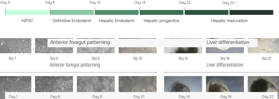
Generation Process
Our liver organoids faithfully replicate the structure and function of human liver tissue by expressing key cellular markers. EpCAM is present in hepatocytes and cholangiocytes, confirming proper epithelial organization. AFP highlights immature hepatocytes, reflecting developmental processes, while Ki67 marks actively dividing hepatocytes and progenitor cells. ZO-1 demonstrates barrier integrity in hepatocytes and cholangiocytes, and HNF4A drives hepatocyte differentiation and maturation. In addition, E-cadherin (ECAD) ensures cell – cell adhesion between hepatocytes and cholangiocytes, supporting robust epithelial structures.
Together, these markers confirm that our liver organoids capture the cellular diversity, architecture, and functional complexity of the human liver – making them a powerful platform for disease modeling, metabolism studies, and drug testing.
Generation of Liver organoids
Cellular and Structural Similarity
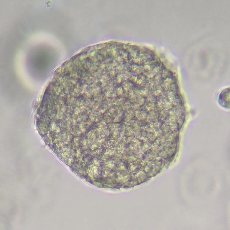
Liver organoids can be observed in a mature form with hepatic layers, bile duct structures architecture similar to actual liver tissue.
They are also composed of various cells such as hepatocytes, cholangiocytes, Kupffer cells, stellate cells.
Expression markers
- EpCAM
Hepatic progenitor - ECAD
Liver epithelial cells - AFP
Immature hepatocyte - HNF4A
Mature hepatocytes - ZO-1
Tight junctions - Ki67
Cell proliferation - FOXJ
Mucous cells
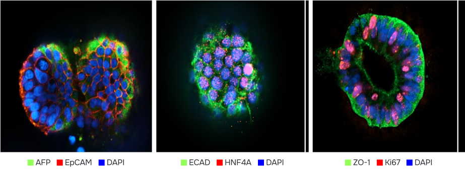
Functional maturation of hLO
Gene expression
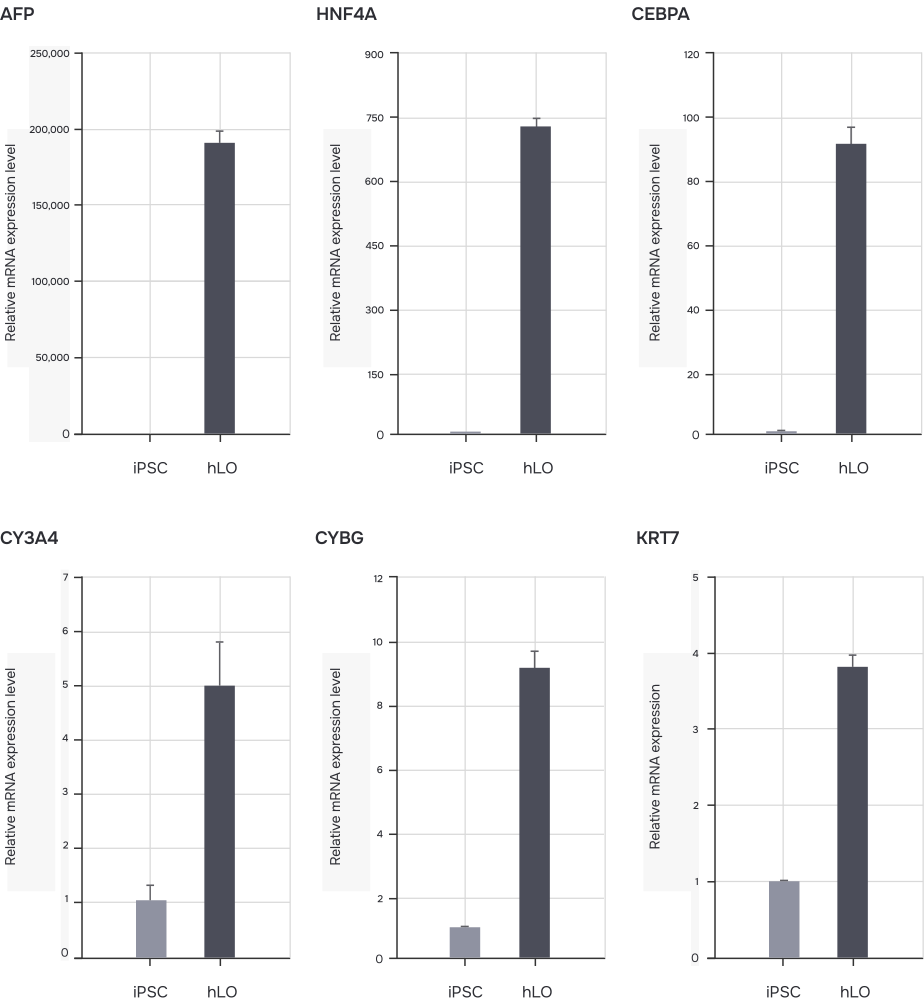
Expression markers
- AFP Immature hepatocyte
- HNF4A Mature hepatocytes
- CEBPA Mature hepatocytes
- CY3A4 Mature hepatocytes
- CYBG Hepatic stellate cells
- KRT7 Biliary epithelial cells (Cholangiocytes)
Fatty Liver Model
Stage of liver disease
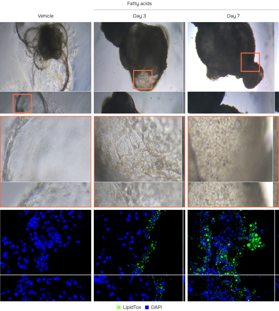
- To induce a fatty liver model, we treated the liver organoids with free fatty acids.
- It was confirmed that markers such as HNF4A, Albumin, and CYP3A4, which are indicative of healthy hepatocyte function, decreased in the damaged model.
- This model can be used to evaluate effective substances related to fatty liver disease treatment.
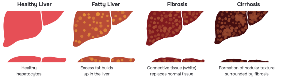
Fatty acid accumulation in hLO