Pulmonary fibrosis is a progressive and irreversible lung disease characterized by excessive extracellular matrix deposition and impaired alveolar regeneration.
Despite decades of research, therapeutic development has been severely constrained by the poor translational relevance of traditional preclinical models. Conventional approaches, including 2D cell cultures and bleomycin-induced animal models, fail to capture the cellular complexity and pathological remodeling of human fibrotic lung tissue.
Key challenge: How can epithelial injury, fibroblast activation, and matrix remodeling be modeled in a human-relevant system that supports translational drug discovery?
Discuss Your Intestinal Fibrosis Research
Our scientists will help design a human-relevant disease modeling study.
Lambda Biologics developed a 3D human lung organoid model that recapitulates key structural and molecular hallmarks of pulmonary fibrosis. The model exhibits fibroblast activation and excessive extracellular matrix deposition, resulting in tissue stiffening and pathological structural remodeling. By reproducing key aspects of fibrotic disease progression, this platform enables quantitative evaluation of antifibrotic candidate compounds in a human-relevant context.
Model highlights:
Human lung organoids
Derived from human lung epithelial progenitor cells
Fibrosis-responsive
Compatible with pro-fibrotic stimulation (e.g. TGF-β)
3D lung architecture
Self-organizing 3D architecture resembling distal lung tissue
Screening-ready
Scalable and reproducible for drug screening applications
Human lung epithelial cells are embedded in a 3D matrix to form stable lung organoids with alveolar-like features.
Step 2: Fibrosis Induction
Organoids are exposed to fibrotic cues (TGF-β, mechanical stress, or fibroblast co-culture) to induce pathological remodeling.
Step 3: Phenotypic & Molecular Readouts
ECM deposition (collagen I, fibronectin)
Myofibroblast activation (α-SMA)
Epithelial integrity and differentiation markers
Transcriptomic and imaging-based analysis
Key Findings
Using this lung organoid platform, we observed:
Robust induction of fibrotic markers following TGF-β stimulation
Structural remodeling resembling early-stage human pulmonary fibrosis
Differential responses to known anti-fibrotic compounds
Improved sensitivity compared to 2D cultures
These results demonstrate the model’s ability to capture disease-relevant biology not accessible in conventional systems.
Model Validation Using a Clinically Approved Antifibrotic Drug
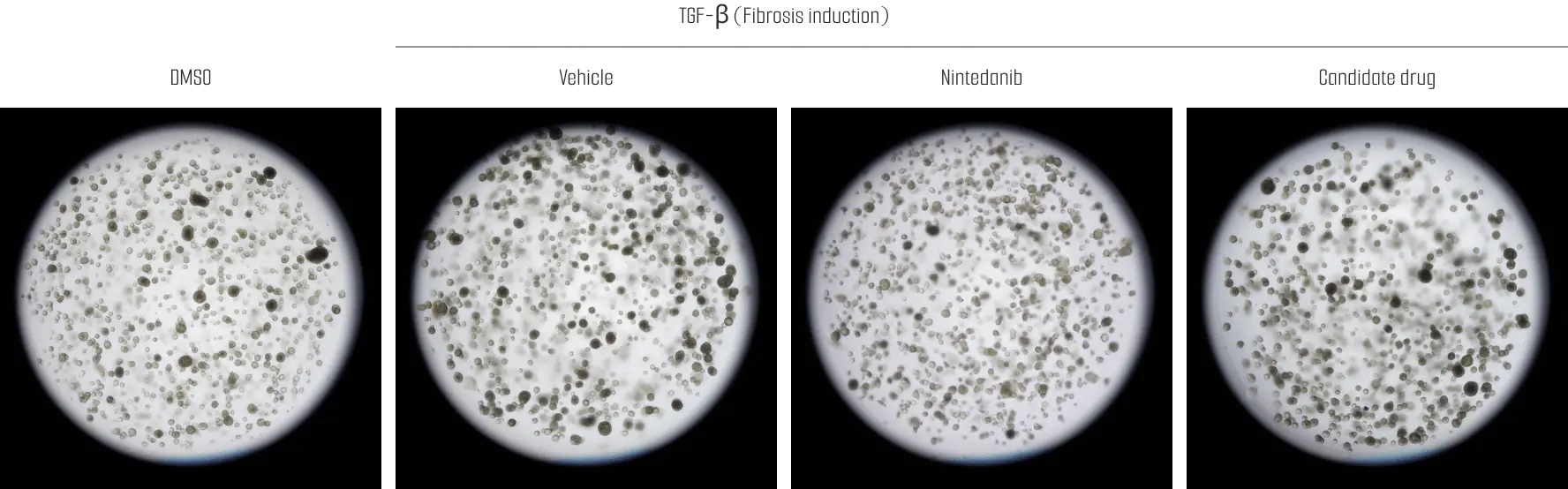
Using a TGF-β–induced fibrosis model in tissue-derived human lung organoids, immunohistochemical analysis revealed that treatment with Nintedanib significantly reduced the expression of fibrosis-associated markers α-SMA and Vimentin, validating the model’s responsiveness to clinically approved antifibrotic therapy.
In addition, multiple candidate compounds elicited comparable reductions in fibrosis marker expression, with certain candidates demonstrating statistically significant effects. These results support the application of this platform for comparative assessment of antifibrotic candidates and early-stage translational decision-making.
Discuss Your Hair Research
Our scientists will help design a human-relevant disease modeling study.